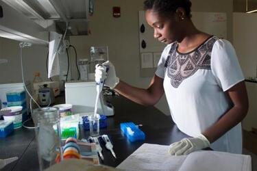
Neuroscience Research and Experiential Learning
August 12, 2016

August 12, 2016
In the lab of Department of Neuroscience Cell Biology & Physiology associate professor David R. Ladle, Ph.D., students study the mechanisms behind lower-limbed movement as a result of changes in neuronal circuitry during development in mouse spinal cords. In order to better understand the goal, students want to look at the spatial organization and connectivity of neuronal groups in the spinal cord over time and relate it to changes in muscle connectivity patterns.
Biological Sciences undergraduate program gratudate (2016) Taylor Floyd (pictured above) studies how a calcium binding protein
Finding the right college means finding the right fit. See all that the College of Science and Math has to offer by visiting campus.